
The scientific study of artists’ materials can answer critical questions about a work’s authenticity, state of preservation, attribution, and restoration history. Exciting discoveries have been made about the palettes and working methods of painters throughout the hundred or more years that scientists have studied works of art. We have recently even gained information about how artists’ masterpieces are being transformed over time by exposure to light and oxygen. While the results of these studies have occasionally been described in the mainstream news, particularly when an infamous forgery or potentially devastating degradation phenomenon is uncovered, rarely can the public examine these findings first-hand.
The spectroscopic signatures of artists’ pigments and paint binding media can look like impenetrable charts or graphs (described by one art lawyer I work with as ‘pages of squiggly lines’, a terminology that broke my heart on the spot). While our blood, sweat, and tears go into extracting these data from artists’ pigments, the spectroscopic signatures of these pigments are readily interpreted only by museum scientists.

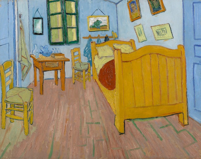
The Bedroom (1888), Vincent Van Gogh. Van Gogh Museum, Amsterdam (Van Gogh Foundation)
Until recently, that is. Within the past 15 years there has been a revolution in how the chemical data on artists’ materials are collected and displayed. Now the latest scientific discoveries on famous artists such as Vincent Van Gogh and Pablo Picasso are available and accessible to a broad community. This includes not only museum professionals, but potentially everyone who is involved in the study of cultural heritage: from art collectors to catalogue-raisonné scholars, art dealers and vetting teams at art fairs. In the last 10 years, X-ray fluorescence technology has enabled the imaging of the chemical elements in numerous paintings, including a Blue period Picasso with a hidden portrait beneath the surface. Most recently, these techniques have been refined by adding X-ray diffraction, enabling the imaging of molecular species within the painting.
For example, Vincent Van Gogh’s bedroom paintings can be chemically imaged to show the location of his Eosin red pigment (or, as he would have called it, ‘geranium lake’). This pigment faded from a bright pink colour to almost white during Van Gogh’s lifetime, often in a matter of months. Scientists are using chemical imaging-techniques to reproduce Van Gogh’s works with their original violet irises that now appear blue, and pink roses that now appear white, allowing the public for the first time to see Van Gogh’s works as he would have painted them. The results are almost as shocking as the reconstructed views of ancient Greek marbles with their original painted surfaces.

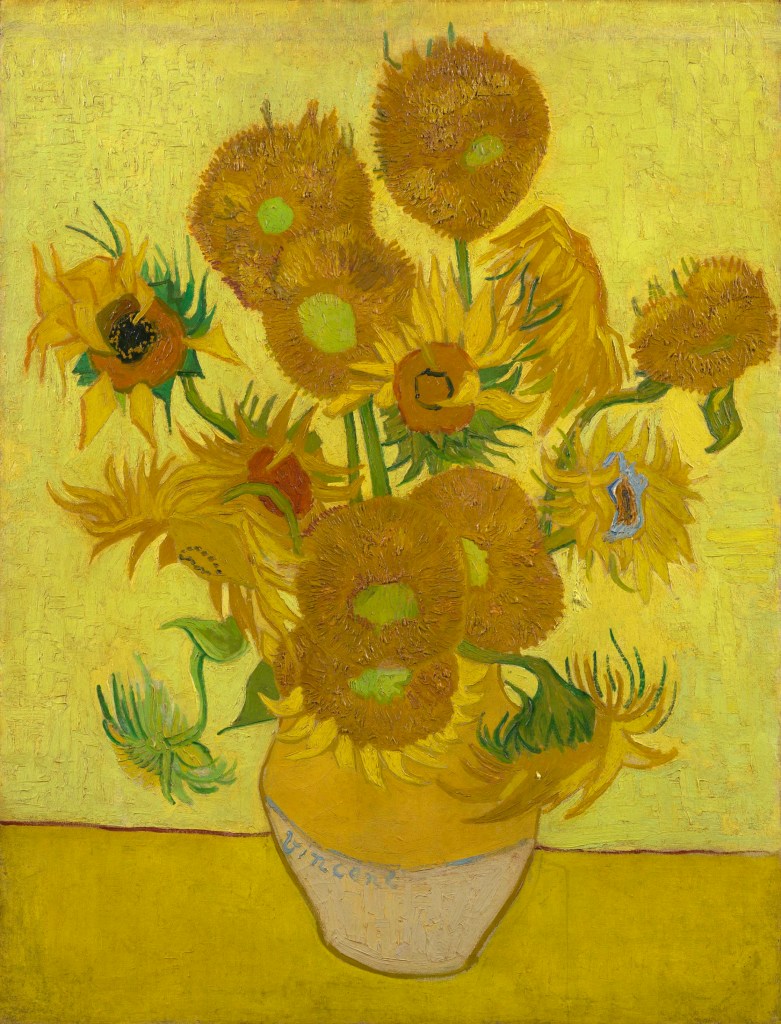
Sunflowers (1889), Vincent Van Gogh. Van Gogh Museum, Amsterdam (Van Gogh Foundation)
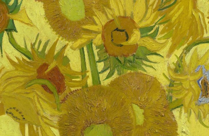
This month’s molecular imaging of Vincent Van Gogh’s Sunflowers (1889), using X-ray diffraction imaging, has revealed the location of the two different forms of chrome yellow used in the painting. Chrome yellow is a lemony-yellow lead chromate pigment that occurs in two different chemical forms, one of which is light-sensitive and the other light-fast. By mapping out the location of these two closely related pigments, researchers have been able to identify the regions of the work that must be closely monitored for colour changes. With over 50 per cent of the yellow paint being prepared from the light-sensitive material, this study demonstrates the care with which this painting must be displayed to prevent future fading and discoloration. As a result, we can see that chemical imaging is vital to the future scientific study of paintings, not only for imaging buried paintings, but also for understanding the original visions of artists.
Jennifer Mass is the Andrew W. Mellon Professor of Cultural Heritage Science at the Bard Graduate Center, New York.