At the end of the 17th century, working in Bologna’s hospital morgues, the Sicilian sculptor Gaetano Giulio Zumbo became the first artist to make anatomical teaching models using coloured wax. The head of an executed criminal, his mouth open and eyes frozen in the final spasm of death, is rendered in wax moulded over a real human skull, the epidermis removed to show the man as machine beneath. Until then, waxworks were largely confined to life-size effigies of saints and anatomical ex-votos, sometimes depicting limbs and organs. The art historian Aby Warburg thought that the individualised portrait that emerged in the Renaissance built on this ‘fetishism of the waxwork cult’. In the 18th century wax began to be used by artists working in tandem with anatomists for scientific purposes.
Zumbo was employed by Cosimo III, the penultimate Medici Grand Duke of Tuscany, to make a series of miniature theatres that record baroque scenes of plague and syphilis: gruesome cribs that illustrate both the process of putrescence and the consequences of vice. They have the compact horror and seasick hues of Géricault’s The Raft of the Medusa, and were displayed in the Uffizi before being transferred to La Specola, Florence’s wax museum. The Marquis de Sade considered them masterpieces, and praised their powerful and ‘fearful truth’: ‘These scenes of plague appealed to my cruel imagination,’ he wrote in his journal, ‘and I mused, how many persons had undergone these awful metamorphoses thanks to my wickedness?’
The anatomy room, with figures by Ercole Lelli, in Palazzo Poggi, Bologna
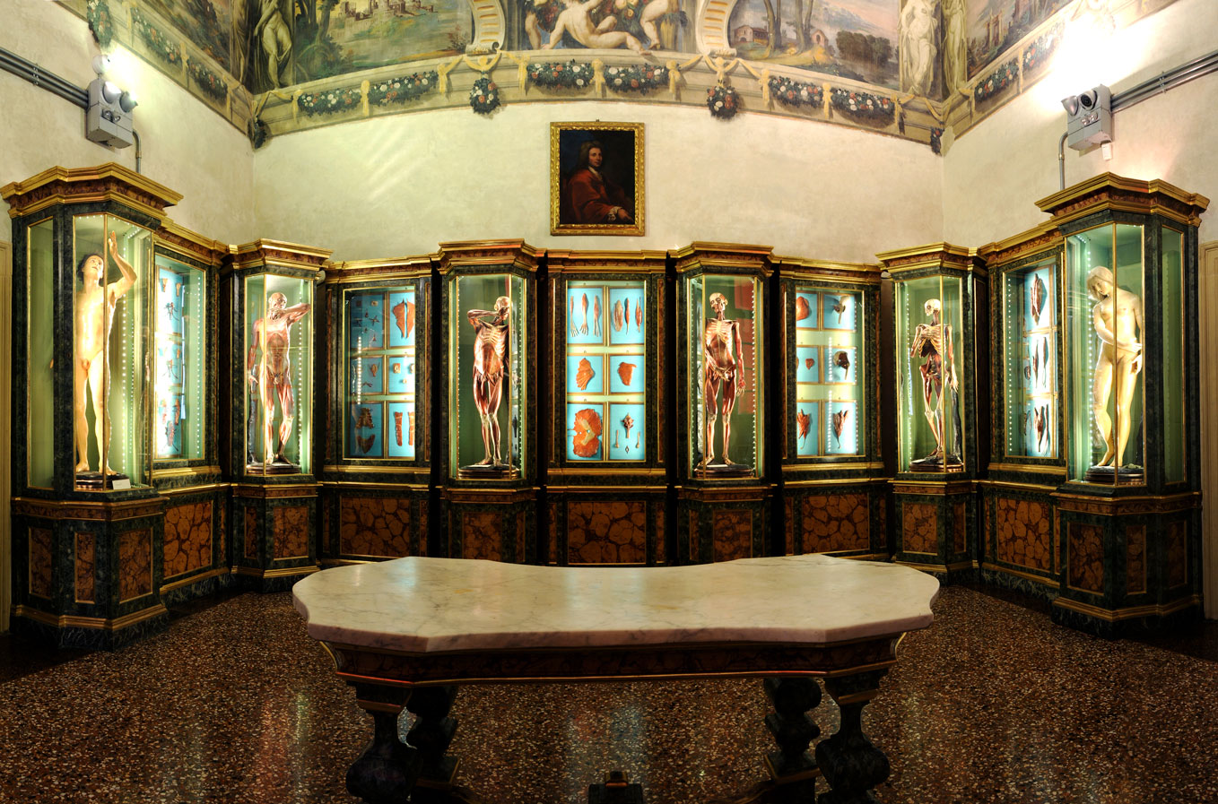
Bologna was the first place in Italy to have a special school of anatomical wax modelling, founded in the 1740s under the patronage of Pope Benedict XIV, almost 50 years after Zumbo practised there. Its head was Ercole Lelli, ‘figure director’ at the art academy located alongside the Institute of Science in Palazzo Poggi. He was commissioned to make eight life-sized statues for the institute’s anatomy room: they depict a naked Adam and Eve, and six other flayed figures, a series in which the wax seemingly melts from real human bone to reveal the raw skeletons beneath, which are shown holding the scythe and sickle of Death. As there was no means of preserving corpses, which made it hard for students to learn directly from cadavers, as many as 200 corpses needed to be dissected to make a single écorché.
Anatomical model (1742–51), Ercole Lelli. Museo di Palazzo Poggi, Bologna

Mounted on green silk in the cabinets between the figures are muscles and organs made of hemp cloth soaked in beeswax mixed with bran and turpentine, the colourful viscera glossed in clear varnish. In this way, the anatomy room brings the anatomical treatises of Leonardo and Michelangelo to life (the Victoria and Albert Museum has some of the latter’s wax maquettes of flayed body parts). Lelli was assisted in the creation of this three-dimensional encyclopaedia of the body by Giovanni Manzolini, who also made wax tablets of the reproductive organs and the evolution of the uterus during pregnancy for the institute’s School of Obstetrics, where midwives were taught to deliver babies through tactile exploration. They practised blindfold using a ‘birthing machine’, a glass ball the size of a belly at full-term, from which they would carefully manipulate a cloth doll.
Bust of Giovanni Manzolini dissecting a heart (1755), Anna Morandi. Museo di Palazzo Poggi, Bologna

Manzolini’s wife, Anna Morandi, who learned wax moulding techniques from her husband, soon exceeded them all in her skill. She was able to manipulate her material to create diaphanous models of insubstantial parts of the body that no one had managed to represent before, including the sensory, urogenital and cardiovascular systems. Morandi displayed them to much acclaim to the Royal Society in London and the court of Catherine the Great in St Petersburg. In Palazzo Poggi the wax portrait busts she created of Manzolini and herself – he dissecting a heart, she a brain – look out on to her almost surrealistic creations. These include a wheel of eyes, each one staring in a different direction, surrounding the starfish-like form of the musculature that controls them; a wax model of her own dexterous hands, one pinching the other; and an outsized ear that could be disassembled to show students its inner workings.
Self-portrait bust dissecting a brain (1755), Anna Morandi. Museo di Palazzo Poggi, Bologna. Photo: Giacinto Cambini/Archivio Opificio delle Pietre Dure di Firenze

Wax modellers from Florence were trained in Bologna, but in the 1770s a rival school of anatomical ceroplastics was established at Florence’s Natural History Museum, now La Specola. Palazzo Poggi has one of several copies of Clemente Susini’s so-called Medici Venus (a title that reflects the fact that La Specola has its origins in that illustrious family’s collection, but also puns on medico, the Italian word for doctor), which was the artistic star of that institution, making it an essential stopping point on any Grand Tour. She lies on her bed in a sensual pose, her neck choked in pearls, and can be dissected or demounted piece by piece with sadistic theatricality. The Medici Venus is housed, like a slashed sleeping beauty, in a glass coffin, and is herself a casket: the layers of her insides have been removed, and placed neatly around her, to reveal a fully formed foetus in the womb, of which there is otherwise no external clue.
Napoleon dispersed the collection when he moved the University of Bologna’s campus to Palazzo Poggi, and the objects were returned only in 2000 when the anatomy rooms were restored. Much of the diaspora had been stored in the Luigi Cattaneo Anatomical Wax Museum, five minutes walk away, which is crammed full of 19th-century waxworks by Giuseppe Astorri and Cesare Bettini that show an intellectual shift from the study of anatomy to pathology. Like Morandi’s wax ear, many of these are enlarged models, and show congenital malformations, such as conjoined twins, facial deformities such as Proteus syndrome, and infectious diseases such as leprosy and smallpox as if seen under a microscope. By the end of the 19th century, photography began to replace wax models, which were increasingly viewed as kitsch curiosities. The work of these highly skilled artist-anatomists was consigned to Chambers of Horrors.
From the December 2020 issue of Apollo. Preview the current issue and subscribe here.